

Multiscale Microstructural Brain Imaging
* This page is only updated periodically (last update 1/18/2025), please see our Publications for most recent work.
We are broadly interested in applying imaging methods for microstructural imaging of the brain. This has applications ranging from basic science such as understand fiber tracts in the brain, to clinical application such as surgical guidance for cancer resection. Primarily, we aim to use polarization-sensitive optical imaging techniques for assessment of cellular structure, anisotropy, and plaque content in brain tissues. This research effort is highly collaborative with Dr. Elizabeth Hutchsinon, who is an expert in brain imaging using magnetic resonance imaging (MRI), which together we aim to combine with optical imaging to yield multiscale information.
Polarization-sensitive imaging, or polarimetry, is a unique tool to understand neural tissue because the myelin sheath that coats neural fibers is intrinsically birefringent. Birefringence is the property of a material to introduce a geometry-dependent phase delay to light (Figure 1). By measuring this phase delay and its spatial orientation, the alignment of white matter fiber tracts can be mapped by imaging with polarization-sensitive systems. Mueller Matrix polarimetry is a comprehensive measurement technique that characterizes the complete polarization response of a material, including birefringence, diattenuation and depolarization. While microstructural orientation characteristics of the tissue can be discerned by probing birefringence, other characteristics tied to the scattering of light can be measured via depolarization, and geometry-dependent absorption through diattenuation.

Fig 1. Graphic visualization of the anisotropic phase delay induced by myelin, causing linearly polarized light to transform into circularly polarized light.
Recent Work
In one research effort, our lab utilizes a full Mueller Matrix Polarimeter developed by Nikon which operates in a backscattering configuration. This microscope captures pixelwise Mueller Matrices across five different imaging wavelengths which can then be decomposed into individual polarization properties of a material (Fig 2). One of the major goals of our work with this system is to develop techniques for imaging neural tissue in reflectance mode to enable point-of-care application, such as with surgical guidance.

Fig 2. (A) Photograph of Mueller Matrix polarimetry system, (B) sectioned ferret brain sample with region of interest showing curved white matter tract, (C) resulting images of the regions of interest, decomposed into polarization properties using the Lu-Chipman decomposition.
Our group has conducted several studies where we imaged various regions of interest in fixed ferret brain tissue in order to assess the sensitivity and response of backscattering polarimetry to tissue structures in white and grey matter [1]. We also have investigated the use of tissue phantoms to determine overall system sensitivity and dependence on non-tissue properties such as object geometry [2]. Recently, we have begun to investigate the application of polarization-sensitive optical coherence tomography (PS-OCT) to yield depth-resolved polarization information (Fig. 3) [3].
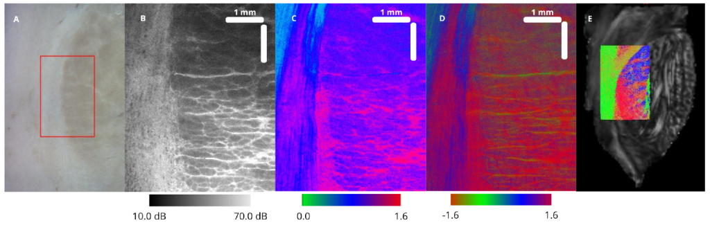
Fig 3. (A) Brightfield photograph of the sample and selected ROI for PS-OCT imaging. (B) PSOCT
Average Intensity measured in dB. (C) PS-OCT Retardance ranging from 0 to π. (D) PS-OCT
Retardance Angle ranging from −π to π. (E) dMRI image of the brainstem and tractography, where
the color coding indicates red for left-right, green for front-back, and blue for up-down directions.
Complementing our optical techniques, we also utilize other imaging technologies such as diffusion MRI with our collaborators. With this data, we have developed advanced co-registration techniques to align these different modalities to evaluate the agreement between the contrast mechanisms between the two imaging modalities [4]. In separate NIH-funded work (R01 943783), we are developing PLI and MRI technology for the purpose of identifying microstructural markers of Alzheimer’s disease pathology [5].
Ongoing and Future Work
Improving methods for neural imaging is desirable across a range of applications. Among them intraoperative surgical guidance and the study of the pathogenesis of neurodegenerative diseases and traumatic brain injuries are some of the most impactful. Advancing this technology towards these applications requires a thorough understanding of the polarization response of tissues and how this response can be altered due to tissue changes. Our future work will focus on categorizing how polarization properties are transformed due to different disease states, ultimately determining whether polarization-sensitive biomarkers have high sensitivity and specificity for identifying disease changes. In addition, further developing polarization-sensitive imaging methods in reflection-based configurations broadly enables in vivo applications of the technique, and we will focus on developing compact and robust point-of-care devices.
References
- Bonaventura J, Morara K, Carlson R, Comrie C, Daigle N, Hutchinson E, and Sawyer TW. Backscattering Mueller Matrix polarimetry on whole brain specimens shows promise for minimally invasive mapping of microstructural orientation features. Front. Photon. 3 (2022).
- Bonaventura J, Morara K, Carlson R, Comrie C, Twer A, Hutchinson E, and Sawyer TW. Evaluating backscattering polarized light imaging microstructural mapping capabilities through neural tissue and analogous phantom imaging. J. Biomed. Opt. 29(5), 05914 (2023).
- Aguilera Cuenca I, Hutchinson EB and Sawyer TW. Estimation and Comparison of Brainstem Fiber Orientation Via Diffusion MRI Tractography and Polarization Sensitive Optical Coherence Tomography. Frontiers in Optics and Laser Science (2024).
- Carlson R, Comrie CJ, Bonaventura J, Morara K, Daigle N, Hutchinson EB, and Sawyer TW. Backscattering Mueller matrix polarimetry estimates microscale anisotropy and orientation in complex brain tissue structure. J. Med. Imag. 12(1), 016001 (2024).
- Comrie CJ, Carlson R, Ahsan Z, Moshkriz A, Sawyer TW, Intorcia A, Serrano GE, Beach TG and Hutchinson EB. Identification of diffusion, kurtosis, and propagator MRI markers of Alzheimer’s Disease pathology in post-mortem human tissue. Imaging Neursci. (2024).