

Weston Welge
 Weston Welge is a fifth-year Ph.D. student in Optical Sciences working with Prof. Jennifer Barton. His research involves the development of optical imaging technology and methods for the detection of cancer. In particular, Weston is working to detect colon cancer using optical coherence tomography (OCT), an imaging modality based on low-coherence interferometry that is capable of generating high-resolution cross-sectional images about 2 mm deep into tissue. While small tumors can be manually identified using OCT, automated detection may be possible by measuring the response of the blood vessels to heat. Healthy blood vessels dilate and increase blood flow when heated, but vessels that develop to support growing tumors are typically unresponsive to heat. The rate of blood flow in the vessels near the tissue surface can be measured using Doppler OCT, a functional form of OCT that directly measures the frequency shift that light experiences when reflecting off of a moving particle. Weston has developed a miniature endoscope for imaging the colon of a mouse model of colon cancer. He is currently studying whether Doppler OCT can detect colon tumors by comparing the change in blood flow rate near the surface of the colon before and after heating by a few degrees. If successful, this method could eventually be used to detect very small pre-malignant tumors that are often missed during colonoscopy.
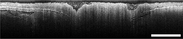
Cross-sectional OCT image of colon adenoma in mouse (scale bar = 1 mm)