Tyler Tate
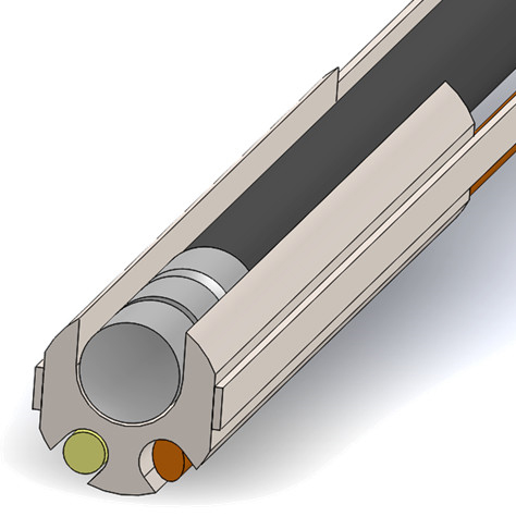
 Tyler Tate works in Dr. Jennifer Barton’s Tissue Optics Laboratory developing endoscopes using optical coherence tomography (OCT) and multi-spectral fluorescence imaging (MFI) to image early stage ovarian cancer. Ovarian cancer is generally treatable if detected early. Unfortunately, current survival rates are very low as ovarian cancer does not typically present symptoms until late stages and no current effective screening currently exists. OCT is the light based analog to ultrasound providing high resolution depth images of tissue microstructure. OCT images have high diagnostic value, but limited field of view not well suited for navigation. MFI provides wide field of view imaging similar to a standard camera. Using ultraviolet light, MFI causes certain proteins and molecules in the body to fluoresce. Imaging the fluorescence provides locations of abnormal molecular concentrations that may indicate developing cancer. MFI then guides the OCT system to abnormal regions for further evaluation. Tyler’s research primarily focuses on combining OCT and MFI into miniature endoscopes to detect early stage cancer in the fallopian tubes and on the ovary. Imaging inside the fallopian tube requires a sub millimeter diameter endoscope that is flexible and steerable. The small optical fibers and lenses necessary to fit both imaging techniques inside the endoscope push the limits of current manufacturing ability. Ultimately the works strives to provide a minimally invasive ovarian cancer screening device.