
Christine Alvarez
I research the light-filtering properties of centric marine diatoms. These diatoms have silica shells with periodic pore structures that act as naturally occuring photonic crystals with a large band gap in the 1-2μm range. We have the ability to both characterize this bandgap and to map out pore spacing across the diatom shell, thus allowing us to correlate pore spacing, location on the diatom valve, and wavelengths of light that are passed through the valve. We are investigating possible biological functions of the photonic properties exhibited by the species C. Wailesii by imaging them under a fluorescence microscope and mapping out both pore and chloroplast location. The amorphous silica in the shell is dyed with Rhodamine and excited with a 408 nm laser such that both the auto-fluorescence of the chloroplasts and the stained shells are visible. In addition, we hope to be able to change diatom pore spacing by cultivating diatoms with various chemicals- specifically, we hope that small concentrations of heavy metal ions will affect pore spacing and size. If we can find a link between water contamination and diatom photonic properties, this could provide a way to test for some aspects of water quality.
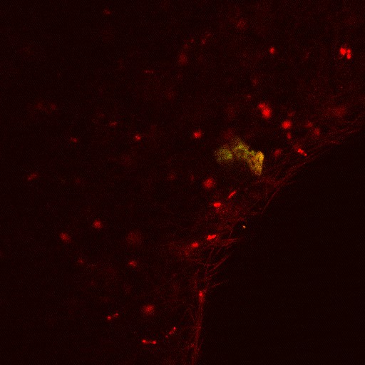
Fluorescence mage of a dyed diatom shell (green/orange) with chloroplasts both within the diatom and from surrounding algae (red).
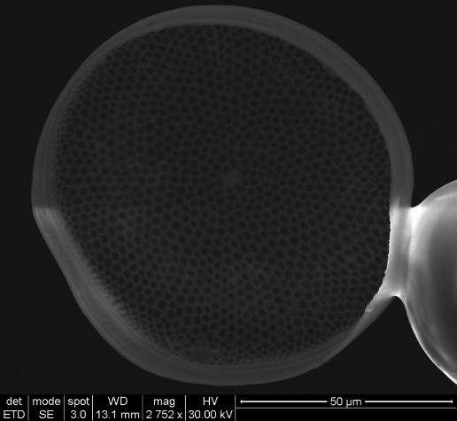
SEM image of a C. Wailesii frustule mounted on a fiber taper. Pores are visible across valve.