Multiphoton Microscopy
Introduction
A multiphoton microscope (MPM) is a device that uses nonlinear optics in order to generate contrast in a sample. These nonlinear optical processes do not require contrast agents like traditional optical microscopes, enabling label-free 3D imaging, determination of diseases, and identification of material properties. One of the main advantages our systems have over existing MPM systems is the use of compact femtosecond fiber lasers, which enable both a small form factor, and also generate a longer excitation wavelength compared to a Ti:Sa laser, enabling deeper tissue penetration. Our systems can use Second Harmonic Generation (SHG), Two photon excitation fluorescence (2PEF), Third Harmonic Generation (THG), and Three Photon Excitation Fluorescence (3PEF) to image samples.
Students: Benjamin Cromey, Ryan Knox, Lam Nguyen, Katarina Korch
Recent Projects
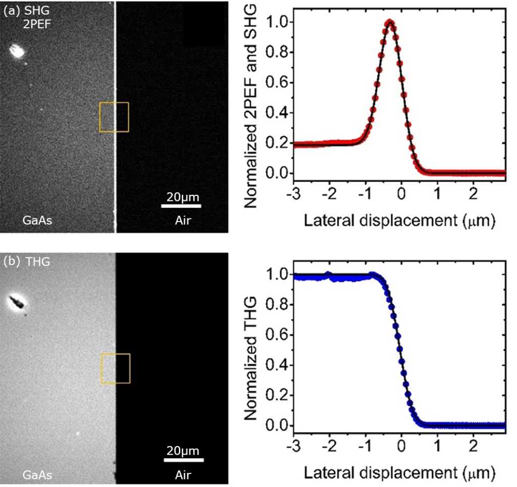
We published a simple yet powerful method for characterizing the performance of a multiphoton microscope using a nonlinear knife-edge technique. This method uses the sharp edge of a Galium Arsenide (GaAs) wafer and several equations to fit the edge profile to produce a final resolution result. Compared to using fluorescent micro-beads, which are prone to damage and photobleach over time, this robust technique can be used under higher irradiance and over longer time without damaging.
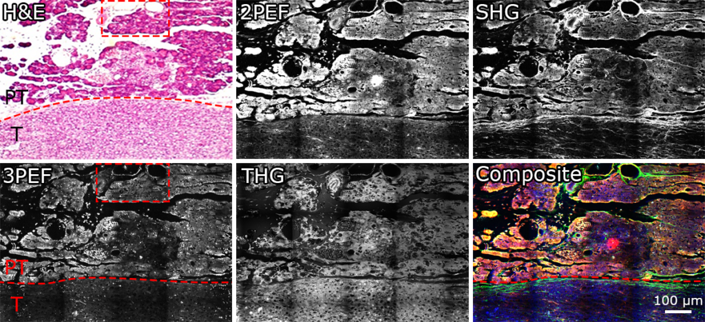
We explored the use of multimodal MPM for the label-free identification of normal and cancerous tissue of the pancreas in a mouse model by comparing the images to H&E microscopy. Our early studies indicate that MPM using second harmonic generation, third harmonic generation and multiphoton excitation of endogenous fluorescent proteins can each contribute to the label-free analysis of the pancreatic surgical margin.
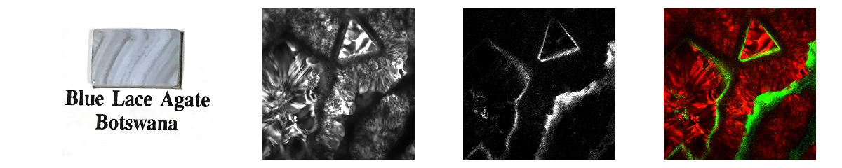
We showed for the first time that the multiphoton microscope can create useful and beautiful 3D images of gems and minerals. We compared our results to images found in the literature with traditional methods of examining stones, and found that we could capture details in 3D that were typically only accessible through sectioning of the minerals. We hope that this is the start of a new research area in gems and minerals. Supplemental images from the paper can be found here. This work was also recently presented at Optics + Photonics in San Diego, and an interview about that talk can be found here.
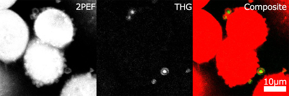
We used targeted microbubbles to help determine the margins of pancreatic cancer. These microbubbles were used to highlight only the cancerous regions of the tissue, with the goal of eventually being used in surgery to greatly assist surgeons by enabling quick and accurate margin determination. Reliably finding the margin between the healthy and cancerous tissue is a crucial part of increasing the chances of survival in this deadly cancer.
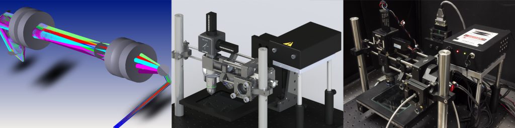
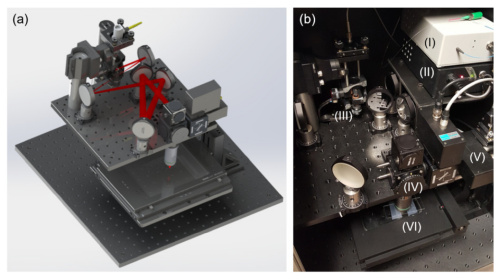
We created a new kind of multiphoton microscope, the first of its kind, an All Reflective Multiphoton Microscope. This mirror based Multiphoton Microscope has the advantage of being a system with no dispersion, enabling the use of any laser source without pulse spreading or chromatic aberrations.
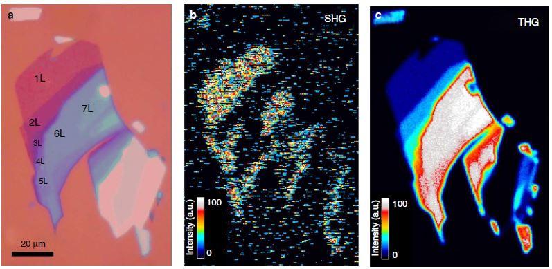
We investigated the nonlinear properties of different layers of Molybdenum Di-sulfide. This material has very strong nonlinear behavior, even creating a detectable Fourth harmonic Generation signal nearly as strong as the second harmonic signal. This nonlinear behavior was explained experimentally as well as theoretically, using a continuum-model Hamiltonian and perturbation theory.