

Resume
- Updated 8-13-17
Navigation
- Areas of Competence
- Educational Background
- Dissertation
- Awards
- Employment History
- Professional Affiliations
- Professional Service
- Proceeding Papers
- Refereed Publications
- Books
- Book Chapters
- Patents
Areas of Competence:
Wavefront sensing, lens design, optical system design and testing, schematic eye modeling, corneal topographic analysis, ophthalmic instrumentation, image quality analysis, aberration theory, Windows programming. Extensive experience with Code V and Zemax lens design software.
Educational Background:
- Ph.D. Optical Sciences, University of Arizona, 1995.
- M.S. Optics, University of Rochester, 1991.
- B.S. Optics, University of Rochester, 1990.
Dissertation:
- Visual Performance Prediction Using Schematic Eye Models.
Awards:
- Innovator of the Year – Academia, Governor’s Celebration of Innovation 2021
- Southern Arizona Innovation Leader of the Year, Southern Arizona Tech + Business Expo 2021
- Tech Launch Arizona I-Squared Awards Inventor of the Year 2020
- Robert R. Shannon Endowed Chair in Optical Sciences 2020
- Leading Edge Research, University of Arizona 2012
- Research to Prevent Blindness Innovative Ophthalmic Research Award 2011
- Research to Prevent Blindness Career Development Award 2002-2006
- Eastman Kodak Fellowship, 1992-1995.
- Eastman Kodak Scholarship, 1987-1990.
Employment History:
College of Optical Sciences, University of Arizona (2010- ). Professor
- Design of variable power, diffractive multifocal and extended depth of field lenses.
- Conventional and Accommodating intraocular lens testing.
- Wavefront sensing, corneal topography, ocular surface metrology and instrument design.
- Computational Photography.
Department of Ophthalmology, University of Arizona (September, 1998-2010). Assistant/Associate Professor/Professor
- Establishment of independent research program based upon wavefront sensing, adaptive optics, corneal topography and schematic eye modeling with applications to refractive surgery, contact lens design, corneal disease detection, assessment of visual performance and the correction of refractive errors.
Optical Sciences Center, University of Arizona (1995-1998). Assistant Research Scientist.
- Developed a keratoconus detection scheme and photorefractive keratectomy analysis package using corneal topographic data.
- Built imaging system for quantifying iris pigmentation.
- Integrated CCD camera into a hand-held direct ophthalmoscope.
- Designed and constructed a stereo-fundus camera.
- Built and tested an operative keratometer for use in refractive surgery.
- Contributed to projects involving the design, fabrication and testing of conformal optics.
Optical Sciences Center, University of Arizona (1991-1995). Graduate Research Associate/Assistant.
- Dissertation research. Modeled human visual system using CODE V lens design software. Predicted visual acuity and change in contrast sensitivity in the presence of refractive error. Analyzed videokeratoscopic corneal height data and applied results to a schematic eye to predict visual performance following refractive surgery.
- Evaluated the performance of novel contact lens design.
- Designed a preliminary lens system for “optical implantation” of intraocular lenses.
- Contributed to projects involving calibration and accuracy analysis of commercially available videokeratoscopes.
- Evaluated the feasibility of an underwater imaging system.
Eastman Kodak Company, Rochester, NY, (Summers 1988-90). Intern.
- Researched coupling infrared laser diodes to thin film waveguides for optical disk applications.
- Examined heating effects of CO2 and Nd:YAG lasers on various film bases.
- Computer-modeled heating effects using a finite element analysis program.
- Tested various schemes for latent image printing, including arrays of Light Emitting Diodes and Liquid Crystal light valves. Evaluated image quality of coherent optical bundle that converted plane waves to cylindrical waves.
Professional Affiliations:
- Optical Society of America (OSA), Fellow
- SPIE, Fellow
- National Academy of Inventors, Senior Member
- Association for Research in Vision and Ophthalmology (ARVO), Silver Fellow
Professional Service:
- Associate Editor of the Journal of Refractive Surgery (2007-2016)
- Publications committee SPIE (2015-2017)
- Refereed manuscripts for numerous optics and ophthalmic journals
Books:
- J. Schwiegerling, Optical Specification, Fabrication and Testing, (SPIE, Washington, 2014).
- M.P Schaub, J. Schwiegerling, E. Fest, R.H. Shepard, A. Symmons, Molded Optics: Design and Manufacture, (Taylor & Francis, Boca Raton, Florida, 2011).
- J. Schwiegerling, Field Guide to Visual and Ophthalmic Optics, (SPIE, Washington, 2004).
Book Chapters:
- J. Schwiegerling. The human eye and its aberrations. In: Handbook of Optical Engineering, 2nd Edition. Ed. Malacara D. (CRC Press, Boca Raton, Florida, 2017).
- J. Schwiegerling. Geometrical optics. In: Handbook of Visual Optics. Ed. Artal P. (Taylor & Francis, Boca Raton, Florida, 2017).
- J. Schwiegerling. Intraocular lenses. In: Handbook of Optics, 3rd Edition. Vol. 3. Ed: Bass, M. (McGraw-Hill, New York, 2010).
- W.A. Maxwell, J. Schwiegerling. Multifocal IOLs: Measuring Aberrations. In: Mastering Refractive IOLs the Art and Science. Ed: Chang D.F. (Slack, New Jersey, 2008).
- Schwiegerling J. The optics of wavefront technology. In: Duane’s Clinical Ophthalmology, Eds: Tasman W, Jaeger EA. (Lippincott, Philadelphia, 2004).
- Schwiegerling J. Wavefront information sampling, fitting and conversion to a correction. In: Wavefront Customized Visual Correction: The Quest for Supervision II. eds. Krueger RR, Applegate RA, MacRae SM. (Slack, New Jersey, 2004).
- J. Schwiegerling, R.W. Snyder and S.M. MacRae “Optical Aberrations and Ablation Pattern Design”, in Customized Corneal Ablation: The Quest for Supervision. ed:S.M. MacRae, R.R. Krueger, R.A. Applegate (Slack, New Jersey, 2001).
Patents:
- J.T. Schwiegerling. “Diffractive Trifocal Lens,” US Patent 11,199,725, Issued December 14, 2021.
- J.T. Schwiegerling. “Diffractive Trifocal Lens,” US Patent 10,725,320, Issued July 28, 2020.
- J.T. Schwiegerling. “Diffractive Trifocal Lens,” US Patent 10,209,533, Issued February 19, 2019.
- J.T. Schwiegerling, N.N. Peyghambarian, G.A. Peyman, N. Savidis. “Holographic Adaptive See-through Phoropter,” US Patent 9,681,800. Issued June 20, 2017.
- J.T. Schwiegerling. “Diffractive Trifocal Lens,” US Patent 9,320,594, Issued April 26, 2016.
- P. Valley, N. Savidis, J.T. Schwiegerling, G. Peyman, N.N. Peyghambarian. “Variable focal length achromatic lens system comprising a diffractive lens and a refractive lens” US Patent 9,164,206, Issued October 20, 2015.
- J.T. Schwiegerling, E. Dereniak, M.W. Kudenov, H. Luo, K. Oka, E.A. DeHoog. “Compact Snapshot Polarimetry Camera,“ US Patent 8,368,889, Issued February 5, 2013.
- J.M. Miller, J.T. Schwiegerling. “Imaging Lens and Illumination System,” US Patent 7,048,379, Issued May 23, 2006.
- J.T. Schwiegerling, W.F. Coyer. “Image Modifiers for use in Scanning Photographic Images” US Patent 6,202,040, Issued March 13, 2001.
Proceedings Papers:
- Moschitta, J. Schwiegerling, “Single-shot intraocular lens surface measurement with the GelSight topography system,” Proc. SPIE 12221, 22211A (2022).
- Su, J. Schwiegerling, “Generalized surface reconstruction and fringe analysis through phase measuring deflectometry,” Proc SPIE 12221, 122210K (2022).
- Cvarch, J. Schwiegerling, “Multifocal Contact Lens HDR Image Simulation Showing Dysphotopsia,” Proc SPIE 12217, 1221704 (2022).
- Sawyers, J. Schwiegerling, “Through focus point spread function and modulus transfer function for multifocal lenses,” Proc SPIE 12217, 1221703 (2022).
- Guan, J. Schwiegerling, “Remote measurement of the clinical prescription of spectacle lenses,” Proc SPIE 12216, 1221606 (2022).
- J. Schwiegerling “Diffractive multifocal lens analysis using complex Fourier series,” Proc. SPIE 12078, International Optical Design Conference 2021, 1207810 (2021).
- J. Schwiegerling, Y. Guan, J. Miller, E. Harvey, “Remote measurement of sphero-cylindrical lens power and orientation through distortion analysis,” Proc. SPIE 11815, 1181502 (2021).
- J. Schwiegerling, “Analysis of extended depth of focus systems with complex pupil decomposition,” Proc. SPIE 11483, 114830D (2020).
- J. Schwiegerling, “The impact of the ocular Shack Hartmann sensor on improving visual performance,” Proc. SPIE 11479, 1148309 (2020).
- J. Schwiegerling, “Rendering light fields for optical system simulation,” Proc. SPIE 11105, 11105E (2019).
- J. Schwiegerling, “Simulating optical system performance using light fields generated from rendering software,” Imaging and Applied Optics, Munich, Germany (2019).
- J. Schwiegerling, “Using light fields to simulate the performance of optical systems,” Proc. SPIE 10743, 1074308 (2018).
- M. Olvera-Angeles, A. Padilla-Vivanco, K. Ortega, J. Sasian, J. Schwiegerling, J. Arines, E. Acosta, “Optimizing trefoil phase plates design for color wavefront coding,” Proc. SPIE 10745, 1074515 (2018).
- J. Schwiegerling, “Image simulation using decomposition of the point spread function,” Proc. SPIE 10690, 1069006 (2018).
- J. Schwiegerling, “Optical transfer function expansion of quadratic pupils,” Proc. SPIE 10590, 1059005 (2017).
- J. Schwiegerling, “Review of Zernike polynomials and their use in describing the impact of misalignment in optical systems,” Proc. SPIE 10377, 103770D (2017).
- J. Schwiegerling, “Linear decomposition of the optical transfer function for annular pupils,” Proc. SPIE 10375, 103750F (2017).
- B. Amirsolaimani, N. Peyghambarian, J. Schwiegerling, A. Bablumyan, N. Savidis, G. Peyman, “An automatic holographic adaptive phoropter,” Proc. SPIE 10352, 1035208 (2017).
- J. Schwiegerling, “Diffraction efficiency and aberrations of diffractive elements obtained from orthogonal expansion of the point spread function,” Proc. SPIE 9953, 995307 (2016).
- Y. Wang, M. Kudenov, A. Kashani, J. Schwiegerling, M. Escuti, “Snapshot retinal imaging Mueller matrix polarimeter,” Proc. SPIE 9613, 96130A (2015).
- J. Schwiegerling, “Optical transfer function optimization based on linear expansions,” Proc. SPIE 9579, 95790H (2015).
- W. J. Duncan, J. Schwiegerling, “Simulating optical system performance with three-dimensional scenes,” Proc. SPIE 9579, 95790F (2015).
- J. Schwiegerling, “Plenoptic camera image simulation for reconstruction algorithm verification,” Proc. SPIE 9193, 91930V (2014).
- J. Schwiegerling, “History of the Shack Hartmann wavefront sensor and its impact in ophthalmic optics,” Proc. SPIE 9186, 91860U (2014).
- J. Schwiegerling, J. S. Tyo, “Relating transverse ray error and light fields in plenoptic camera images,” Proc. SPIE 8842, 884203 (2013).
- J. Schwiegerling, “Tolerancing considerations for visual systems,” Proc. SPIE 8491, 849104 (2012).
- J. Schwiegerling, G. C. Birch, J. S. Tyo, “Analysis and compression of plenoptic camera images with Zernike polynomials” Proc SPIE 8487, 84870G (2012).
- G. C. Birch, J. S. Tyo, J. Schwiegerling, “3D astigmatic depth sensing camera,” Proc. SPIE 8129, 812903 (2011).
- P. Valley, M. R. Dodge, J. Schwiegerling, D. Mathine, G. Peyman, N. Peyghambarian, “Flat liquid crystal diffractive lenses with variable focus and magnification,” Proc. SPIE 7786, 77860H (2010).
- G. Li, P. Valley, P. Ayräs, J. Haddock, M. S. Giridhar, D. Mathine, J. Schwiegerling, G. Meredith, B. Kippelen, S. Honkanen, N. Peyghambarian, “High-efficiency switchable diffractive lens,” Proc. SPIE 6310, 63100H (2006).
- B. E. Bagwell, D. V. Wick, R. Batchko, J. D. Mansell, T. Martinez, S. R. Restaino, D. M. Payne, J. Harriman, S. Serati, G. Sharp, J. Schwiegerling, “Liquid crystal based active optics,” Proc. SPIE 6289, 628908 (2006).
- J. Andrews, S. Teare, S. Restaino, C. Wilcox, D. Wick, H. Xiao, J. Schwiegerling, “Dynamic aberration control testbed for the characterization of multiple wavefront sensors,” Proc. SPIE 6018, 60180R (2005).
- J. Andrews, S. Teare, S. Restaino, C. Wilcox, D. Wick, H. Xiao, J. Schwiegerling, “Optical testbed for comparative analysis of wavefront sensors,” Proc. SPIE 5892, 589221 (2005).
- J. Straub, J. Schwiegerling, “Surgical and healing changes to ocular aberrations following refractive surgery,” Proc. SPIE 4951, (2003).
Refereed Publications:
- Akhoundi, E. Ozgur, C. Draper, R. Voorakanam, J. Wycoff, D. Reetz, P.-A. Blanche, L. Lacomb, G. Peyman, J. Schwiegerling, N. Peyghambarian, “Performance analysis of a compact auto-phoropter for accessible refractive assessment of the human eye,” Appl. Opt. 61, 2207-2212 (2022)..
- Lapid-Gortzak C. Bala, J. Schwiegerling, R. Suryakumar, “New methodology for measuring intraocular lens performance using acuity reserve,” J. Cataract Refract. Surg. 47, 1006-1010 (2021).
- McAlinden, J. Schwiegerling, J. Khadka, K. Pesudovs,” Corneal aberrations measured with a high-resolution Scheimpflug tomographer: repeatability and reproducibility,” J. Cataract Refract. Surg. 46, 581-590 (2020).
- E. DeHoog, R. Van Dine, L Fitzgerald-DeHoog, J. Schwiegerling, “Relating wavefront error to visual acuity in pre and post-LASIK eyes: a comparison of methods,” J. Opt. Soc. Am. A 37, 192-198 (2020).
- M. Olvera-Angeles, A. Padilla-Vivanco, J. Sasian, J. Schwiegerling, J. Arines, E. Acosta, “Effect of spherical aberration in trefoil phase plates on color wavefront coding,” Jpn. J. Appl. Phys. 57, 08PF05 (2018).
- S.J. McCafferty, E.T. Enikov, J. Schwiegerling, S.M. Ashley, “Goldmann tonometry tear film error and partial correction with a shaped applanation surface,” Clin. Ophthalmol. 12, 71-78 (2018).
- S. McCafferty, J. Levine, J. Schwiegerling, E.T. Enikov, “Goldmann and error correcting tonometry prisms compared to intracameral pressure,” BMC Ophthalmol. 18, 2 (2018).
- S. McCafferty, J. Levine, J. Schwiegerling, E.T. Enikov, “Goldmann applanation tonometry error relative to true intracameral intraocular pressure in vitro and in vivo,” BMC Ophthalmol. 17, 215 (2017).
- B. Amirsolaimani, G. Peyman, J. Schwiegerling, A. Bablumyan, N. Peyghambarian, “A new low-cost, compact, auto-phoropter for refractive assessment in developing countries,” Sci Rep. 7, 13990 (2017).
- M. Vinas, C. Dorronsoro, A. Radhakrishnan, C. Benedi-Garcia, E.A. LaVilla, J. Schwiegerling, S. Marcos, “Comparison of vision through surface modulated and spatial light modulated multifocal optics,” Biomed. Opt. Express. 8, 2055-2068 (2017).
- S. McCafferty, G. Lim, W. Duncan, E.T. Enikov, J. Schwiegerling, J. Levine, C. Kew, “Goldmann tonometer error correcting prism: clinical evaluation,” Clin Ophthalmol. 11, 835-840 (2017).
Summary: Modified tip of Goldmann tonometer that gives a more precise measure of intraocular pressure - J. Schwiegerling, “Relating wavefront error, apodization, and the optical transfer function: general case,” J. Opt. Soc. Am. A 34, 726-731 (2017).
Summary: Creates a novel linear expansion for the OTF where the expansion coefficients can be related to the wavefront error coefficients for both on- and off-axis cases. - S. McCafferty, G. Lim, W. Duncan, E. Enikov, J. Schwiegerling, “Goldmann Tonometer Prism with an Optimized Error Correcting Applanation Surface,” Transl Vis Sci Technol. 5, 4 (2016).
Summary: Modified tip of Goldmann tonometer that gives a more precise measure of intraocular pressure - L. Werner, J.C. Stover, J. Schwiegerling, K.K. Das, “Light scattering, straylight, and optical quality in hydrophobic acrylic intraocular lenses with subsurface nanoglistenings.” J Cataract Ref Surg 42, 148-156 (2016).
Summary: Light Scatter, MTF and Badal images are assessed in explanted lenses with nanoglistenings - A.H. Mahamat, F.A. Narducci, J. Schwiegerling. “Design and optimization of a volume-phase holographic grating for simultaneous use with red, green and blue light using unpolarized light.” Appl Opt 55, 1618-1624 (2016).
Summary: Coupled-wave theory is used to develop a Bragg grating with high diffraction efficiency at multiple visible wavelengths - R.F. Steinert, J. Schwiegerling, A. Lang, A Roy, K. Holliday, E. Barragán Garza, A.S. Chayet. “Range of refractive independence and mechanism of action of a corneal shape-changing hydrogel inlay: Results and theory.” J Cataract Refract Surg 41, 1568-1579 (2015).
Summary: Determines the method of action for a small corneal inlay which provides extended depth of focus - J.M. Miller, E.M. Harvey, J. Schwiegerling. “Higher-order aberrations and best-corrected visual acuity in Native American children with a high prevalence of astigmatism.” J AAPOS 19, 352-357 (2015).
Summary: Examines higher order aberrations and visual acuity in a population with high astigmatism - S.J. McCafferty, J.T. Schwiegerling. “Deformable Surface Accommodating Intraocular Lens: Second Generation Prototype Design Methodology and Testing.” Transl Vis Sci Technol 4.2.17 (2015).
Summary: Describes a prototype accommodating intraocular lens that changes power with surface deformation - S.C. Cole, L. Werner, J. Schwiegerling, A. Crandall. “Visual aberrations in a multifocal intraocular lens with injection-related scratches.” J Cataract Ref Surg 40, 1913-1918 (2014).
Summary: Examines the optical performance of an damaged IOL with scratches - J. Schwiegerling. “Relating wavefront error, apodization, and the optical transfer function: on-axis case.” J Opt Soc Am A 31, 2476-2483 (2014).
Summary: Creates a novel linear expansion for the OTF where the expansion coefficients can be related to the wavefront error coefficients - E. Swan, J. Schwiegerling, G. Peyman, E. Enikov. “Photostress testing device for diagnosing retinal disease.” Photonics 1, 211-219 (2014).
Summary: Describes a prototype device for photostress testing - E.M. Harvey, J.M. Miller, J. Schwiegerling. “Utility of an open field Shack-Hartmann aberrometer for measurement of refractive error in infants and young children.” J AAPOS. 17, 494-500 (2013).
Summary: Validates the performance of a custom built handheld open view Shack Hartmann sensor - J. Schwiegerling. “Eye axes and their relevance to alignment of corneal refractive procedures.” J. Refract. Surg. 29, 515-516 (2013).
Summary: Reviews the various definitions of the axes of the eyes - N. Savidis, G. Peyman, N. Peyghambarian, J. Schwiegerling. “Nonmechanical zoom system through pressure-controlled tunable fluidic lenses.” Appl. Opt. 52, 2858-2865 (2013).
Summary: Demonstrates a zoom system with fixed fluidic lenses - E.M. Harvey, J.M. Miller, J. Schwiegerling, D. Sherrill, D.H. Messer, V. Dobson. “Developmental changes in anterior corneal astigmatism in Tohono O’odham Native American infants and children.” Ophthalmic Epidemiol. 20, 102-108 (2013).
Summary: Changes in astigmatism with age in a Native American population - K.K. Das, J.C. Stover, J. Schwiegerling, M. Karakelle. “Technique for measuring forward light scatter in intraocular lenses.” J. Cataract. Refract. Surg. 39, 770-778 (2013).
Summary: Measurement of forward light scatter in intraocular lenses. - S. J. McCafferty, J.T. Schwiegerling, E.T. Enikov. “Thermal load from a CO2 laser radiant energy source induces changes in corneal surface asphericity, roughness, and transverse contraction.” Invest. Ophthalmol. Vis. Sci. 53, 4279-4288 (2012).
Summary: Changes in corneal shape induced by laser heating. - S. J. McCafferty, J.T. Schwiegerling, E.T. Enikov. “Corneal Surface Asphericity, Roughness, and Transverse Contraction after Uniform Scanning Excimer Laser Ablation.” Invest. Ophthalmol. Vis. Sci. 53, 1296-1305 (2012).
Summary: Examines morphological change in corneal shape and surface roughness following exposure with excimer laser pulses - G.C. Birch, J.S. Tyo, J. Schwiegerling. “Depth measurements through controlled aberrations of projected patterns.” Opt. Express 20, 6561-6574 (2012).
Summary: Measures a 3D depth map of a scene by projecting a structured light pattern that various with distance. - E.M. Harvey, J.M. Miller, J. Schwiegerling, C.E. Clifford-Donaldson, T.K. Green, D.H. Messer, V. Dobson. “Accuracy and validity of IK4 handheld video keratometer measurements in children.” J. AAPOS 15, 407-409 (2011).
Summary: Measures the accuracy of a handheld keratometer against a commercially available system. - J. Schwiegerling, “Scaling pseudo-Zernike expansion coefficients to different pupil sizes,” Opt Lett. 36, 3076-3078 (2011).
Summary: Illustrates a simple method for rescaling a Pseudo-Zernike expansion to smaller concentric sub-apertures. - J.A. Davison, A.S. Patel, J.P. Cuhna, J. Schwiegerling, O. Muftuoglu, “Recent studies provide an updated clinical perspective on blue light-filtering IOLs,” Graefes Arch. Clin Exp. Ophthalmol. 249, 957-968 (2011).
Summary: Analyzes recent clinical and laboratory data to demonstrate capabilities of blue light filtering IOLs. - P. Valley, N. Savidis, J. Schwiegerling, M.R. Dodge, G. Peyman, N. Peyghambarian, “Adjustable hybrid diffractive/refractive achromatic lens,” Opt. Express 19, 7468-7479 (2011).
Summary: Examines the achromatic a hybrid electro active diffractive lens combined with a fluidic refractive lens. - E.M. Harvey, V. Dobson, J.M. Miller, J. Schwiegerling, C.E. Clifford-Donaldson, T.K. Green, D.H. Messer, “Prevalence of corneal astigmatism in Tohono O odham Native American children 6 months to 8 years of age,” Invest. Ophthalmol. Vis. Sci. 52, 4350-4355 (2011).
Summary: Tracks the prevalence of astigmatism in a Native American group through early childhood. - P. Valley, M.R. Dodge, J. Schwiegerling, G. Peyman, N. Peyghambarian, “Nonmechanical bifocal zoom telescope,” Opt. Lett. 35, 2582-2584 (2010).
Summary: Demonstrates a non-mechanical zoom system based on pairs of electro-active diffractive lenses. - J. Schwiegerling, E. DeHoog. “Problems testing diffractive intraocular lenses with Shack–Hartmann sensors,” Appl. Opt. 49, D62-D68 (2010).
Summary: Investigates the wavelength dependence and spot splitting that occurs when testing diffractive IOLs with a Shack Hartmann sensor. - R. Marks, D.L. Mathine, G. Peyman, J. Schwiegerling, N. Peyghambarian. “Adjustable adaptive compact fluidic phoropter with no mechanical translation of lenses,” Opt. Letters 35, 739-741 (2010).
Summary: Demonstrates a system for correcting the spherocylindrical error of the eye with a set of fluidic lens. - P. Valley, D.L. Mathine, M.R. Dodge, J. Schwiegerling, G. Peyman, N. Peyghambarian. “Tunable focus flat liquid-crystal diffractive lens,” Opt. Letters 35, 336-338 (2010).
Summary: Demonstrates a liquid crystal Fresnel zone plate with adjustable discrete foci. - J. Schwiegerling. “Predicting clinical visual acuity of presbyopic treatments,” J. Refract. Surg. 26, 66-70 (2010).
Summary: Examines correlating clinical visual acuity with through-focus MTF data obtained with the defocus transfer function. - W. Peng, E. DeHoog, J. Schwiegerling. “Systematic error of a large dynamic range aberrometer,” Appl. Opt. 48, 6324-6331 (2009). Appl. Opt. 48, 6376-6380 (2009).
Summary: Explores systematic errors induced by large defocus levels and their correction in a Shack Hartmann wavefront sensor. - W. Peng, S. Liu, E. DeHoog, J. Schwiegerling. “Systematic errors analysis for a large dynamic range aberrometer based on aberration theory,” Appl. Opt. 48, 6324-6331 (2009).
Summary: Examines the aberrations induced by relaying the Shack Hartmann spot pattern onto a detector from the theoretical standpoint. - R. Marks, D.L. Mathine, J. Schwiegerling, G. Peyman, N. Peyghambarian. “Astigmatism and defocus wavefront correction via Zernike modes produced with fluidic lenses,” Appl. Opt.48, 3580-3587 (2009).
Summary: Testing of properties and aberrations associated with fluidic lenses that are capable of correcting both sphere and cylinder error. - R. Marks, D.L. Mathine, G. Peyman, J. Schwiegerling, N. Peyghambarian. “Adjustable Fluidic Lenses for Ophthalmic Corrections,” Opt. Lett. 34, 515-517 (2009).
Summary: Fluidic lenses that are capable of correcting both sphere and cylinder error are demonstrated. - J. Schwiegerling. “Statistical Generation of Normal and Post-refractive Surgery Wavefronts,” Clin. Exp. Optom. 92, 223-226 (2009).
Summary: A technique for creating wavefront aberration coefficients that are statistically consistent with a given population is demonstrated. - J. Schwiegerling, C. Paleta –Toxqui. “Minimal Movement Zoom Lens,” Appl. Opt. 48, 1932-1935 (2009).
Summary: Two sets of Alvarez lenses are used to create a Keplarian telescope. A 4x magnification is demonstrated with minimal lateral motion of the plates. - E. DeHoog, J. Schwiegerling. “Fundus Camera Systems: A Comparative Analysis,” Appl. Opt.48, 221-228 (2009).
Summary: Fundus camera with internal and external illumination channels are analyzed. - E. DeHoog, J. Schwiegerling. “Optimal Parameters for Retinal Illumination and Imaging in Fundus Cameras,” Appl. Opt. 47, 6769-6777 (2008).
Summary: The illumination channel for a fundus camera is designed and optimized. - J. Schwiegerling, J. Choi. “Application of the Polychromatic Defocus Transfer Function to Multifocal Lenses,” J. Refract. Surg. 24, 965-969 (2008).
Summary: The spectral properties of the optical transfer function and the photopic response of the eye are used to extended the Defocus Transfer Function to the polychromatic case. - J. Choi, J. Schwiegerling. “Optical Performance Measurement and Night Driving Simulation of ReSTOR, ReZoom, and Tecnis Multifocal Intraocular Lenses in a Model Eye,” J Refract Surg 24, 218-222 (2008).
Summary: Night driving scenes for various multifocal IOLs are analyzed - P. Jain, J. Schwiegerling. “RGB Shack-Hartmann Wavefront Sensor.” J. Mod. Optics 4-5, 737-748 (2008).
Summary: Red, green and blue lasers are used as sources in a Shack Hartmann sensor. A color camera is used to capture and separate the resulting spot patterns to measure aberrations at three wavelengths simultaneously. - J. Schwiegerling. “Analysis of the Optical Performance of Presbyopia Treatments with the Defocus Transfer Function,” J. Refract. Surg. 23, 965-971 (2007).
Summary: The effect of diffractive and zonal refractive IOLs as well as apodizing filters on Visual Performance is explored with the defocus transfer function. - E.J. Sarver, J. Schwiegerling, R.A. Applegate. “Extracting Wavefront Error from Shack-Hartmann Image Using Spatial Demodulation,” J. Refract. Surg. 22, 949-953 (2006).
Summary: Fourier techniques are used to recover wavefront error. - J.L. Beverage, J. Schwiegerling. “A Shack-Hartmann-based Autorefractor,” J. Refract. Surg. 22, 932-937 (2006).
Summary: A traditional Shack-Hartmann wavefront sensor has been simplified for measurement of refractive error. - G. Li, D.L. Mathine, P. Valley, P. Ayras, J.N. Haddock, M.S. Giridhar, G. Williby, J. Schwiegerling, G.R. Meredith, B. Kippelen, S. Honkanen, N. Peyghambarian. “Switchable Electro-Optic Diffractive Lens with High Efficiency for Ophthalmic Applications,” Proc. Natl. Acad. Sci. 103, 6100-6104 (2006).
Summary: Development of a liquid crystal Fresnel lens for a switchable bifocal spectacle lens. - J. Schwiegerling, “Blue-Light Absorbing Lenses and Their Effect on Scotopic Vision,” J. Cataract Ref. Surg. 32, 141-144 (2006).
Summary: Analyzes the change in scotopic sensitivity caused by the Alcon Natural (blue-light absorbing) intraocular lens. - J. Schwiegerling, “Recent Developments in Pseudophakic Dysphotopsia,” Curr. Opin. Ophthalmol. 17, 27-30 (2006).
Summary: Examines recent findings and theories of positive and negative dysphotopsia attributed to intraocular lens edge designs. - J. Schwiegerling, “Modal Reconstruction Methods with Zernike Polynomials,” J. Refract. Surg. 21, S552-S557 (2005).
Summary: Compares different techniques for reconstructing wavefronts with Zernike polynomials and examines an example of keratoconus detection. - J. Schwiegerling, “Gaussian weighting of ocular wavefront measurements,” J. Opt. Soc. Am. A 21, 2065-2072 (2004).
Summary: Incorporates a gaussian apodization filter in the pupil and optimizes the degree of apodization to correlation weighted wavefront error with measures of visual performance. - J. Schwiegerling, “Scaling Zernike Expansion Coefficients to Different Pupil Sizes,” J. Opt. Soc. Am. A. 19, 1937-1945 (2002).
NOTE: There is an error in this paper. In Table 1, the expression for b1m should read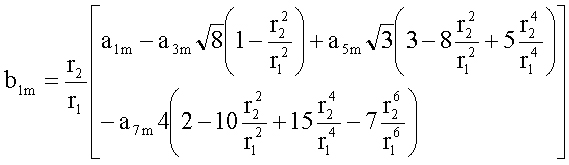
Also, there is a typo in equation A8 of the appendix. The exponent on the very last r1 in equation A8 should be |m|+2i and not |m|-2i.
Summary: Provides simple formulas for converting Zernike expansion coefficients to different pupil sizes.
- J. Schwiegerling, R.W. Snyder, J.H. Lee, “Wavefront and Topography: Keratome-induced Corneal Changes Demonstrate that Both are Needed for Custom Ablation,” J. Refract. Surg. 18, S584-588 (2002).
Summary: Keratome incisions of the cornea alter the aberration structure of the eye. This paper demonstrates some techniques for extracting these changes form topography and aberrometry data. - J.M. Miller, R. Anwaruddin, J. Straub, J. Schwiegerling, “Higher Order Aberrations in Normal, Dilated, Intraocular Lens, and Laser In Situ Keratomileusis Corneas,” J. Refract. Surg. 18, :S579-83 (2002).
Summary: Compares spherical aberration in normal, LASIK and pseudophakic eyes. - J.M. Miller, J. Schwiegerling, H.L. Hall, T. Surachatkumtonekul, “Detection of Improper Fixation in MTI Photoscreening Images,” J. AAPOS 5, 35-43 (2001).
Summary: The effects of fixation error on measurements with the MTI photoscreener are assessed. Trained observers can miss small but relevant errors in fixation. - J. Schwiegerling, “Theoretical Limits to Visual Performance,” Surv. Ophthalmol. 45,139-146 (2000).
Summary: Analysis of a diffraction-limited eye model with chromatic aberration and Stiles-Crawford effect to examine theoretical limits to grating acuity. - J. Schwiegerling, R.W. Snyder, “Eye Movement during Laser In Situ Keratomiluesis,” J. Cataract Refract. Surg. 26, 345-351 (2000).
Summary: A video-based technique for examining eye motion during refractive surgery procedures is outlined. Several examples are shown to illustrate mean and standard deviation of the pupil center relative to the laser axis. - S.M. MacRae, J. Schwiegerling, R. Snyder, “Customized Corneal Ablation and Super Vision,” J. Refract. Surg. 16, S230-S235 (2000).
Summary: Review of emerging technologies for performing customized corneal ablations. - J. Schwiegerling and R. W. Snyder, “Corneal Ablation Patterns to Correct for Spherical Aberration in Photorefractive Keratectomy,” J. Cataract Refract. Surg. 26, 214-221 (2000).
Summary: Pre- and post-operative corneal topographies of PRK patients are analyzed to show and increase in the induced spherical aberration caused by treatment with the Summit OmniMed laser. The induced spherical aberration is shown to be linearly proportional to the degree of myopic correction. - J. Schwiegerling and R.W. Snyder, “Optical Issues in Keratoplasty Patients,” Operative Techniques Cat Ref Surg. 2, 85-88 (1999).
Summary: Basic introduction into some of the optical phenomenon such as aberrations, decentration and scatter that occur in penetrating keratoplasty patients. - J. Schwiegerling and R.W. Snyder, “Visual Performance Modeling Following RK, PRK, and Lasik Demonstrates the Need for Improved Treatment Algorithms,” Ophthalmic Prac. 17, 66-70 (1999).
Summary: Discusses methods for customizing schematic eye models with corneal topography and generating a map of refractive error as a function of position in the entrance pupil. Compares refractive error maps for non-surgical and post-refractive surgery patients to illustrate the introduction of spherical aberration. - S. MacRae, J. Schwiegerling and R.W. Snyder, “Customized and Low Spherical Aberration Corneal Ablation Design,” J. Refract. Surg. (suppl) 15, S246-S248 (1999).
Summary: Analysis of customized schematic eye models of PRK patients to determine the effects of the refractive surgery on the optical properties of the eye. Low spherical aberration ablations were generated for these patients. - J. Schwiegerling and R.W. Snyder, “Custom Photorefractive Keratectomy Ablations for the Correction of Spherical and Cylindrical Refractive Error and Higher Order Aberrations,” J. Opt. Soc. Am. A, 15, 2572-2579 (1998).
NOTE: There is a typo in this paper. Equation (18) should readM’ = SR + CR/2 + (fxpre + fypre)/2Summary: A formal treatment of the shape of a general PRK ablation pattern which corrects for spherical refractive error, astigmatism at any orientation and higher order “spherical-like” aberration. The article considers the patient’s pre-operative corneal topography, refraction and spherical aberration levels to generate an appropriate ablation pattern. - J. Schwiegerling, “Cone Dimensions in Keratoconus Using Zernike Polynomials,” Optom. Vis. Sci., 74, 963-969 (1997).
Summary: TMS-1 corneal topographic height data from clinically diagnosed keratoconus patients is reviewed. Each set of corneal height data is decomposed into Zernike polynomials. The spherical and astigmatic components of the cornea are subtracted to reveal the cone. The location of the cone, its lateral dimensions and its height above the base cornea are determined. The results show that the location of the cone is not at the location of peak dioptric power. This technique may prove useful for tracking the progression of the cone. - J. Schwiegerling and J.E. Greivenkamp, “Using Corneal Height Maps and Polynomial Decomposition to Determine Corneal Aberrations,” Optom. Vis. Sci., 74, 906-916 (1997).
Summary: The use of corneal topographic height data as a complement and/or alternative to dioptric power data is reviewed. Methods for viewing height data and analyzing corneal shape are examined. Advantages of decomposition into orthogonal sets of polynomials is discussed. - J. Schwiegerling and J.E. Greivenkamp, “Keratoconus Detection Based on Videokeratoscopic Height Data,” Optom. Vis. Sci., 73, 721-728 (1996).
Summary: TMS-1 corneal topographic height data from normal and clinically diagnosed keratoconus patients are compared. Each set of corneal height data is decomposed into Zernike polynomials. Two expansion coefficients are shown to be elevated in keratoconic patients and are used in a disease detection method. This detection scheme is compared to other methods such as elevated SAI values and the Rabinowitz I-S calculation. - J. Schwiegerling, J.E. Greivenkamp, J.M. Miller, R.W. Snyder and M.L. Palmer, “Optical Modeling of Radial Keratotomy Incision Patterns,” Am. J. Ophthalmol., 122, 808-817 (1996).
Summary: TMS-1 corneal topographic height data from 8 incision RK patients is obtained. Each set of corneal height data is decomposed into Zernike polynomials and polynomial orders lower than 8-fold symmetry are removed from the original corneal height data. The residual height shows a spoke-like variation in the cornea resulting from the incisions. The optical effects of these “spokes” is analyzed by applying the pattern to the cornea of a schematic eye model. The effects of optical zone size and procedure centration on contrast sensitivity are analyzed. This paper also introduces a schematic eye model which mimics clinically measured values of spherical and chromatic aberration. - J.E. Greivenkamp, M.D. Mellinger, R.W. Snyder, J.T. Schwiegerling, A.E. Lowman and J.M. Miller, “Comparison of Three Videokeratoscopes in Measurement of Toric Test Surfaces,” J. Refract. Surg., 12, 229-239 (1996).
Summary: Toric test surfaces with 1.00, 3.00, 5.00 and 7.00 diopters of astigmatism are used to examine the accuracy of three commercially available corneal topographers. The systems examined are the Computed Anatomy TMS-1, the EyeSys topographer and the Alcon EH-270. Levels of accuracy are comparable between the three devices with an rms power error of approximately 0.25 D and an rms height error on the order of several microns. - J. Schwiegerling and J.E. Greivenkamp, ” Visual System Modeling: Putting the Pieces Together,” in “Optics in 1995,” Opt. and Phot. News 6, 36-37 (December 1995).
Summary: Review of techniques for performing schematic eye modeling and including the effects of the retina and brain processing. - J.E. Greivenkamp and J. Schwiegerling, ” Modeling Soft Contact Lenses in Raytrace Code,” OSA Engineering & Laboratory Notes, Opt. and Phot. News 6 (1995) and Appl. Opt. 34, 8076-8077 (1995).
Summary: Describes a novel technique for incorporating soft contact lenses into raytracing code. We used the interferogram file found in Code V to model decentration effects of soft contact lenses. - J. Schwiegerling, J.E. Greivenkamp and J.M. Miller, “Representation of Videokeratoscopic Height Data with Zernike Polynomials,” J. Opt. Soc. Am. A, 12, 2105-2113 (1995).
Summary: A thorough description of using Zernike polynomials to analyze corneal height data. Gram-Schmidt methods for decomposing corneal height data into Zernikes is described. Conversion of expansion coefficients into clinically familiar terms such as spherical and astigmatic dioptric power and axis are given. Examples of astigmatism, keratoconus and RK height maps are shown. - J.E. Greivenkamp, J. Schwiegerling, J.M. Miller and M.D. Mellinger, ” Visual Acuity Modeling Using Optical Raytracing of Schematic Eyes,” Am. J. Ophthalmol., 120, 227-240 (1995).
Summary: A technique for predicting visual acuity from schematic eye models is described. The technique combines exact raytracing of a schematic eye model with the retinal and brain function to find a visual acuity. Modeling predictions for pupil sizes ranging from 0.5 mm to 8 mm and refractive errors from 0.00 diopters to -5.00 diopters are compared to clinical measurements. High correlation is found between the model predictions and clinical findings.